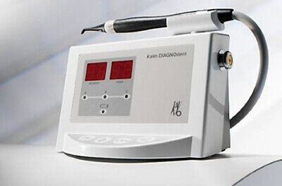
The DIAGNOdent™ will show where the decay lies. It works quickly and reliably. The laser fluorescence detector within the DIAGNOdent is a precise method for identifying fissure caries, proximal caries and periodontitis.
Sub-surface caries lesions can be extremely difficult to detect using an explorer, and the DIAGNOdent™ offers a perfect adjunct to the diagnostic arsenal. It is especially useful for pit and fissure areas – even when the outer tooth surface seems to be intact.
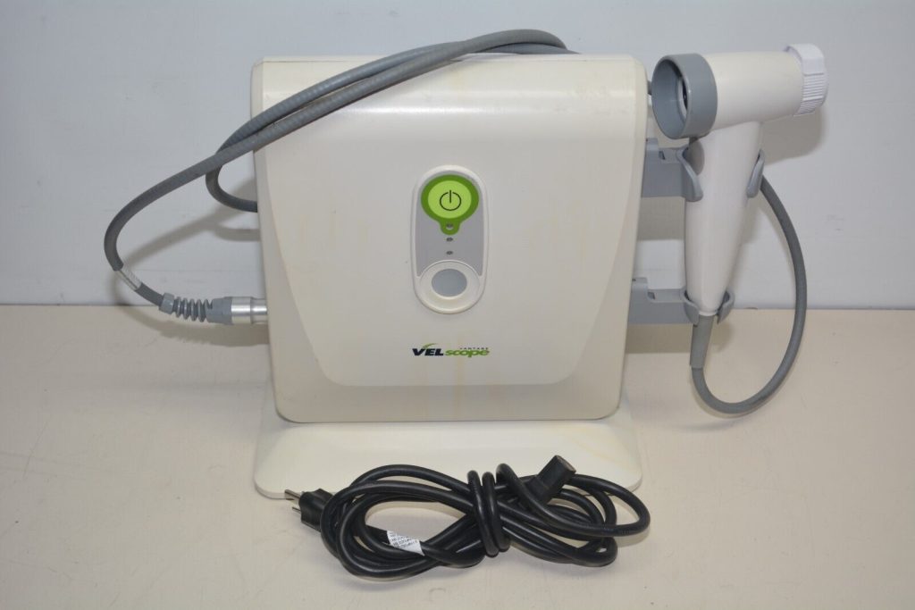
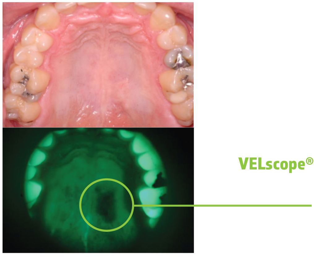
The VELscope system is a powerful, fluorescence-based device that enhances the visualization of oral mucosal abnormalities such as oral cancer and premalignant dysplasia. Unlike other adjunctive devices used by dentists, the VELscope system does not require any dyes or prolonged testing procedures. In fact, VELscope examinations can be performed in the dentist’s office during routine hygiene exams.
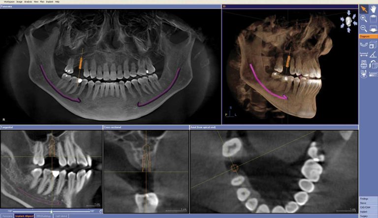
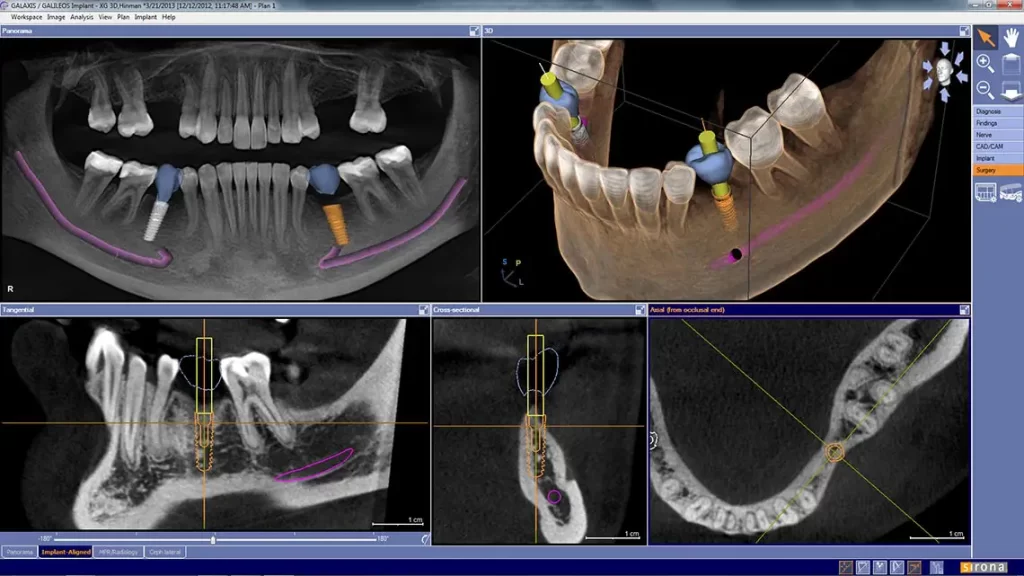
Calismiles is now offering advanced three-dimensional dental imaging to provide patients with the best treatment possible.
This dental imaging technology is particularly useful for dental implant treatment planning, root canal therapy, TMJ and other complex procedures, offering a quick scan that improves the opportunity to catch issues early.
This scanning process takes only 14 seconds and provides detailed images that increase clinical safety. The doctor will review the scan with the patient, using the rotating and moving image capabilities to enhance visibility.
By using this advanced technology, patients can easily see their teeth, upper and lower jaws, sinuses, and any issues that need immediate attention.
Overall, this 3D dental imaging system provides reliable and accurate results that ultimately improve patient care and satisfaction.
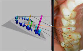
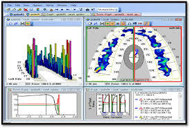
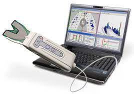
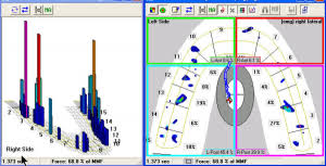
T-Scan is the complete digital occlusal analysis solution helps clinicians to identify premature contacts, high forces, and interrelationship of occlusal surfaces. This important data cannot be captured by traditional, analog occlusal methods, like articulating paper. Whether eliminating destructive forces on a new restoration, or performing an occlusal analysis and adjustment procedure, T-Scan helps balance patients’ occlusions and reduce costly repeat visits and remakes |
